
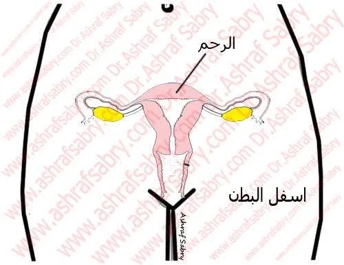
The female reproductive system is a group of organs responsible for conception and pregnancy. It lies in the lower abdomen and pelvis and has internal and external parts.
Struggling to conceive? If natural conception is difficult in your case, IVF and ICSI with Dr. Ashraf Sabry may be the right solution. Learn about the steps, timing and who it suits.
Internal reproductive organs
The vagina
The vagina is a muscular canal about 10 cm long, running from the vulva to the uterus. Its inner lining has folds. Cells, glands and beneficial bacteria produce acidic secretions that keep it moist and protect it from harmful microbes. The vagina stretches during intercourse, and at ejaculation sperm are deposited at its upper end.
The uterus
The uterus is a hollow, pear-shaped muscular organ in the lower abdomen, above the vagina. It provides the passage for sperm on their way to the egg, and later receives and nourishes the fertilized egg. It has two parts: the body of the uterus and, below it, the cervix. The body is about 7 cm long, 5 cm wide and weighs about 50 g, varying between women and with previous pregnancies. In about 80% of women the uterus tilts forward towards the abdominal wall (anteverted), and in about 20% backwards towards the spine (retroverted), which is a normal variant. The uterus is lined by the endometrium, which thickens each month in response to female hormones so a fertilized egg can implant; if pregnancy does not occur, the extra lining is shed as the period.
The cervix
The cervix is the gateway at the lower end of the uterus through which sperm enter. Its lower part can be seen at the top of the vagina, while its upper part lies above it. It is about 3 cm long, with an external opening leading to the cervical canal and an internal opening into the body of the uterus. The canal has folds lined with mucus-producing cells. The mucus changes through the cycle: scanty and thick after a period, then abundant, clear and stretchy in the days before ovulation to help healthy sperm through, then scanty and thick again after ovulation to block sperm. Inside the canal, the mucus protects sperm from vaginal acidity and nourishes them.
The fallopian tubes
The two fallopian tubes are narrow tubes about 10 cm long, one on each side of the uterus, ending in finger-like fringes (fimbriae). The part nearest the ovary, the ampulla, is wider than the part nearest the uterus. When an ovary releases an egg, the fimbriae pick it up and draw it into the tube. The ampulla is where sperm meet and fertilize the egg; the fertilized egg is then carried towards the uterus by rhythmic muscle contractions and by tiny hair-like cilia lining the tube, whose cells also secrete fluids that nourish it.
The ovaries
The two ovaries are small organs on either side of the pelvis, near the ends of the fallopian tubes. They are the source of eggs and of female hormones, making them central to fertility. Each is about 3 cm long and 1.5 cm wide, with an outer cortex containing follicles at different stages and an inner medulla containing blood vessels and nerves. A baby girl is born with about one to two million follicles; around 300,000–400,000 remain at puberty, and the number keeps falling, especially after 35.
Primordial follicles
A primordial follicle consists of an egg surrounded by a layer of granulosa cells; those closest to the egg are called corona cells. Each month, hormones recruit a group of follicles, of which usually one reaches maturity and is ready for fertilization.
External genitalia (the vulva)
The mons pubis
A pad of fatty tissue over the pubic bone, covered with hair after puberty.
The labia majora
Two thick outer folds of skin on either side of the vaginal opening, covered with hair after puberty and containing the openings of some glands.
The labia minora
Thinner inner folds of skin that run alongside the vaginal opening, inside the labia majora.
The clitoris
A small structure at the top of the labia minora, equivalent to the penis in men. It is made of erectile tissue that fills with blood during arousal and has a very rich nerve supply, playing a key role in sexual arousal. It is covered by a fold of skin called the clitoral hood.
The hymen
A thin rim of tissue around the vaginal opening, with an opening that allows menstrual blood to pass. Its shape varies widely between women. It may stretch or tear at first intercourse, but not always, and its appearance is not a reliable indicator of previous intercourse.
Bartholin’s glands
Two glands on either side of the vaginal opening that secrete mucus for lubrication during intercourse.
The urethral opening
The opening of the urethra lies just below the clitoris, above the vaginal opening.


International Guidelines
In line with global standards, Dr. Sabry follows the latest recommendations from the American Society for Reproductive Medicine (ASRM), European Society of Human Reproduction and Embryology (ESHRE), and Royal College of Obstetricians and Gynaecologists (RCOG). Thus, patients receive care that is safe, ethical, and evidence-based.
Our Patient Reviews
Discover what patients say about us on Google Maps, Vezeeta, and WhatClinic.
Why Choose Dr. Ashraf Sabry?
Dr. Ashraf Sabry, consultant in obstetrics, gynecology, fertility, and aesthetic gynecology, is recognized among the best OBGYN and fertility specialists in Cairo. With over 20 years of experience, he combines science and compassion to provide world-class care. Book your appointment through the contact page, WhatsApp, or call +201022224442.